
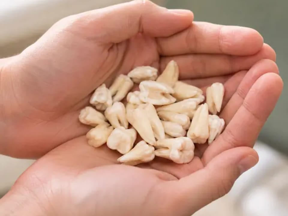
A wisdom tooth breaks through only halfway. The gum settles for a week, then flares again. Food keeps finding the same awkward pocket behind the second molar. These stop-start symptoms are easy to postpone, yet the real position of the tooth remains hidden. With us, an X-ray is used to see where the crown, roots and neighbouring structures actually lie.
Does Every Third Molar Have to Go?
No. Some wisdom teeth erupt with enough room and behave no differently from the molar in front: they meet the bite, stay free from decay and do not defeat the toothbrush. Such a tooth can be left alone and checked during routine visits.
Trouble usually develops in cycles. A flap of gum becomes sore for the second or third time (pericoronitis). Decay appears where a filling cannot be placed reliably. The tooth leans against the second molar, or an image reveals a cystic change around its crown. An abscess is a more urgent version of the same story.
The decision about wisdom tooth removal should follow an examination and imaging. “It might cause trouble one day” is not the same as finding a present clinical reason to operate.
Impacted, Partly Erupted or Sideways
An impacted third molar remains partly or completely beneath gum or bone. A displaced tooth points in an unfavourable direction—towards the second molar, towards the cheek or almost horizontally along the jaw.
During impacted wisdom tooth removal, the surgeon may need to open the gum and clear a small amount of bone over the crown. The tooth can then be divided. Taking it out in sections often requires less force than trying to deliver the whole thing through a narrow opening.
This is what patients usually mean by difficult or surgical wisdom tooth extraction. The name sounds dramatic; the X-ray is more useful. It shows whether the roots are separate, hooked, fused or lying close to a structure that must be protected.
These features often occur in the same tooth, but they describe different things:
| Tooth | What it means |
|---|---|
| Impacted | Remains partly or completely beneath the gum or bone. For removal, the surgeon may need to open the gum, clear a small amount of bone over the crown and divide the tooth. |
| Displaced (dystopic) | Points in an unfavourable direction: towards the second molar, towards the cheek or almost horizontally along the jaw. |
Why Lower Wisdom Teeth Need Careful Mapping
A sensory nerve runs through a canal inside the lower jaw. In some patients, the roots of the eighth tooth sit close to it. The nerve carries feeling from the lower lip and chin, so its relationship with the roots matters before lower wisdom tooth extraction.
A panoramic X-ray gives the first view. If the overlap looks close, cone beam CT may be requested for a three-dimensional map. The scan does not make the operation necessary; it helps the surgeon choose a safer route when treatment is already indicated.
Upper wisdom teeth present a different puzzle. Their roots may be near the maxillary sinus. Again, anatomy decides the plan.
What Happens During the Operation?
First comes local anaesthetic and a short wait. The area is tested. Pressure, movement and vibration may remain noticeable, but a sharp feeling means the dentist pauses.
For an erupted tooth, wisdom tooth extraction may resemble an ordinary removal. A tooth still buried under gum or bone needs access. The surgeon exposes the crown, removes only the bone required for a working path and separates the tooth when that reduces resistance.
After all parts are accounted for, the socket is checked and the gum is repositioned. Dissolving stitches may be placed. Gauze pressure helps the first clot settle over the wound.
Some procedures are quick. A horizontal lower third molar with curved roots is not a quick version of the same job—it is a different piece of surgery.
Swelling, Soreness and the Week Ahead
Swelling often grows during the first days. That does not automatically mean infection. The cheek can feel firm, the jaw may open less freely and chewing may be tiresome for several days.
After that early peak, the general trend should turn. Less pressure. Easier opening. A little more comfort each day. The stiffness should ease gradually; if the mouth opens less and less easily or the stiffness does not improve, contact the dentist.
Protect the clot on day one: no hard rinsing, repeated spitting or socket inspection with a finger. Eat after the numbness has gone and start with soft, lukewarm food.
That post-surgery cigarette is particularly unhelpful. Suction can disturb a young clot, while smoke works against the tissue trying to close the wound. Vaping does not solve either problem. Give the socket a genuine break.
Watch the direction of recovery rather than counting every sore hour. A cheek that is slowly softening is reassuring. Pain that suddenly begins drilling towards the ear after a few calmer days—or swelling that carries on enlarging—belongs back in the dental chair. A foul taste or smell strengthens the reason to call.
